Stem Cell Therapy
Biobest have been involved in the culture of mesenchymal stem cells since 2005 and have provided cells for more than 1,600 stem cell treatments for horses and dogs to date.

Biobest are the largest VMD authorised Stem Cell Centre currently operating in the UK. We culture stem cells directly from bone marrow aspirates or adipose tissue, these can then be used in clinical applications, primarily the treatment of tendon and ligament injuries.
- The use of mesenchymal stem cells for the treatment of tendon and ligament injuries is based on over 10 years of research carried out at the Royal Veterinary College.
- A growing number of small animal practices are performing stem cell treatment in dogs citing joint problems as the main target for the therapy, especially those conditions that end up as arthritis, such as hip dysplasia, elbow dysplasia and osteochondrosis.
- Recent publications have shown that stem cell treatment halves the re-injury rate when compared with conventional therapies.
- Biobest use QC checks at each stage of the process to ensure that the client receives the highest quality cells. Prior to release cells are checked for sterility, viability and the number of cells dispatched is confirmed.
- Biobest’s experience in both producing and sending stem cells for many years allows us to receive and dispatch cells ready to implant in fully validated temperature controlled packaging systems at ambient temperature. We believe this is superior to sending frozen cells in unlicensed chemicals such as DMSO.
- Biobest also work in partnership with Nupsala Veterinary Services who can provide advice, training and support for veterinary orthopaedic treatments.
Stem Cell Forms
Click here to download stem cell submission forms
Stem Cell Therapy - Consumables
Biobest provide aspiration kits containing all the consumables required to take a sample. The sample is then sent to us and we isolate and culture the stem cells in our dedicated cell culture laboratory. Once the required number of cells have been cultured we will contact the practice directly to arrange transport of the cells for implantation. Cells are returned to customers suspended in the patients own plasma, this avoids the need to use unlicensed cryoprotectants and cell culture reagents in a therapeutic product. Our cells are shipped in fully validated ‘biotherm’ boxes to ensure that they reach the practice in top condition.
Stem Cell Therapy - Cryopreservation of Cells
As well as sending stem cell suspensions for implantation we can store cultured cells in liquid nitrogen for future use. In order to ensure safe storage all of our cultures are split between two locations. Cells are held in vessels which are monitored in realtime to ensure optimal storage conditions. Our cryosorage vessles are all alarmed with staff on call 24/7 to deal with any emergencies. If required we can quickly resuscitate the cells from cryostorage and send them out for use, thus preventing the need for further bone marrow aspirates. Bone marrow aspirates can also be taken at the same time as other surgical procedures and stored for use in the event of future injury. Cryopreserved cells can typically be dispatched to a practice for implantation within 7-10 days of resuscitation.
In November 2019 we paid a visit to ICR Vets in Loanhead to speak with vet Chris Sawyer to discuss his experience administering stem cell therapy to a four year of Labrador with osteoarthritis, using stem cells cultured at Biobest.
The video contains some before and after footage of the animal which show the kind of improvement stem cell therapy can deliver.
The Process
The first step in the process is to obtain a bone marrow sample. Bone marrow is a good source of mesenchymal stem cells which can be used for soft tissue repair. The sample is taken by the vet using standing sedation, there is no need for general anaesthesia. Bone marrow is typically taken from either the sternum or tuber coxae of the injured animal. Biobest have recently been granted approval for the use of adipose tissue as an alternative source of mesenchymal stem cells.
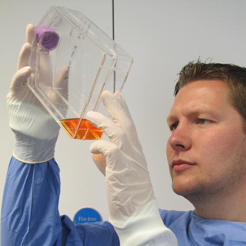
The sample is then sent to Biobest for processing. On arrival at the laboratory our team of cell culturists examine the sample to ensure that there has not been any deterioration in transit. The bone marrow is then centrifuged to separate it into its constituent parts. The part which is richest in stem cells is transferred to a tissue culture flask and over the next few days the stem cells will attach to the flask and start growing. After 7-10 days the cells will usually have reached a stage where they are ready to be split into larger flasks so we can obtain the required number of cells for implantation. Cells will typically be ready to dispatch after 3-4 weeks.
Prior to release a number of QC checks are performed to ensure the cells are sterile, viable and the correct number of cells are present. Cells are then packaged in a validated transport container to minimise degradation during transit and are returned to the practice for delivery the following day. The vet then implants the cells back into the lesion and the horse can begin a rehabilitation programme.
About Stem Cells
Stem cells are unique in that they have the capacity to develop into a range of specialized cell types. This fact has made them a very attractive target for regenerative therapies. Stem cells can be broadly split into 2 types, embryonic stem cells which have the capacity to differentiate into any cell type and adult stem cells which are more limited in the cell types they can become. Using bone marrow aspirates we can isolate mesenchymal stem cells which have the capacity to differentiate into tendon cells, bone cells, cartilage and fat and as such are perfect candidates for the regeneration of soft tissue damage. Mesenchymal stem cells can also be isolated from other tissue, for example, adipose tissue or umbilical cord.
As long ago as 1961 it was shown that mesenchymal stem cells (MSC’s) could develop into tendons in the lab and by the late 1990’s the regeneration of tendon-like tissue had been shown in vivo. Post-mortem results from MSC treated suspensory branches show good longitudinal orientation of fascicle and a collagenous matrix which exhibits a crimp pattern characteristic of ligament rather than scar tissue. Post-mortem results from MSC treated superficial digital flexor tendons show very good tendon healing and a fascicular arrangement that is largely retained / reconstituted and contains an adequate longitudinally arranged fibroblastic / tenocytic population.